- صفحه نخست |
- سفارش کتاب |
- چاپ کتاب |
- فروشگاه |
- اخبار |
- درباره ما |
- ارتباط با ما |
- عضویت در سایت |
- ورود به سایت |
- ویدئوها
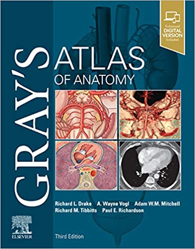
جزییات کتاب Gray's Atlas of Anatomy 2021
طبقه : آناتومی
اطلس آناتومی گری
Gray's Atlas of Anatomy 2021
مشخصات کتاب
ISBN (شابک): | 032363639X- 9780323636391 |
قطع: | رحلی |
ناشر: | |
تعداد صفحات: | 648 |
سال و نوبت چاپ: | 3rd Edition-2021 |
نوع جلد: | هارد |
قیمت خرید کتاب : 1,299,200 تومان
جزییات بیشتر :
Clinically focused, consistently and clearly illustrated, and logically organized, Gray's Atlas of Anatomy, the companion resource to the popular Gray's Anatomy for Students, presents a vivid, visual depiction of anatomical structures. Stunning illustrations demonstrate the correlation of structures with clinical images and surface anatomy - essential for proper identification in the dissection lab and successful preparation for course exams.
- Build on your existing anatomy knowledge with structures presented from a superficial to deep orientation, representing a logical progression through the body.
- Identify the various anatomical structures of the body and better understand their relationships to each other with the visual guidance of nearly 1,000 exquisitely illustrated anatomical figures.
- Visualize the clinical correlation between anatomical structures and surface landmarks with surface anatomy photographs overlaid with anatomical drawings.
- Recognize anatomical structures as they present in practice through more than 270 clinical images - including laparoscopic, radiologic, surgical, ophthalmoscopic, otoscopic, and other clinical views - placed adjacent to anatomic artwork for side-by-side comparison.
- Gain a more complete understanding of the inguinal region in women through a brand-new, large-format illustration, as well as new imaging figures that reflect anatomy as viewed in the modern clinical setting.
- Evolve Instructor site with an image and video collection is available to instructors through their Elsevier sales rep or via request at https://evolve.elsevier.com.
از نظر بالینی متمرکز ، مداوم و به روشنی مصور ، و به طور منطقی سازمان یافته ، اطلس خاکستری آناتومی ، منبع همراه با آناتومی محبوب گری برای دانش آموزان ، تصویری زنده و بصری از ساختارهای آناتومیک ارائه می دهد. تصاویر خیره کننده نشان دهنده ارتباط ساختارها با تصاویر بالینی و آناتومی سطح است - برای شناسایی مناسب در آزمایشگاه قطع و آمادگی موفقیت آمیز برای امتحانات دوره.
دانش آناتومی موجود خود را با ساختارهایی ارائه دهید که از یک جهت گیری عمیق سطحی و عمیق ارائه شده و پیشرفت منطقی را از طریق بدن نشان می دهد.
ساختارهای متنوع آناتومیکی بدن را شناسایی کرده و با راهنمایی بصری تقریباً هزار چهره مصور برجسته آناتومیکی ، روابط آنها را با یکدیگر بهتر بشناسید.
ارتباط بالینی بین ساختارهای آناتومیکی و نقاط دیدنی سطح با عکسهای آناتومی سطحی که با نقوش آناتومیک پوشانده شده است را تجسم کنید.
ساختارهای آناتومیکی را از طریق بیش از 270 تصویر بالینی - از جمله لاپاروسکوپی ، رادیولوژی ، جراحی ، چشم ، چشم و سایر دیدگاههای بالینی - که در مجاورت کارهای هنری آناتومیک قرار گرفته اند ، مقایسه کنید.
درک کاملتری از منطقه اینگوینال در زنان از طریق یک تصویر جدید با فرمت بزرگ ، و همچنین چهره های تصویربرداری جدید که منعکس کننده آناتومی در محیط بالینی مدرن هستند ، بدست آورید.
سایت Evolve Instructor با مجموعه عکس و فیلم از طریق نمایندگی فروش Elsevier یا از طریق درخواست در https://evolve.elsevier.com در اختیار مربیان است.
.
خريد کتاب پزشکي با تخفيف, usmle step 1 books, usmle step 2 books, usmle step 3 books, usmle, usmleiran, usmle ایران, مرکز تخصصی usmle در ایران, 2021, 2022, امتحانات کانادا و استراليا و امريکا , آزمون هاي پزشکي خارج از کشور, مديکال بوک, کتاب رفرنس پزشکي, دانلود کتاب پزشکي, انتشارات کتب پزشکي, انتشارات پزشکيبازدید : 2663 مرتبه
محصولات مشابه
ناشرین
Elsevier (679) LWW (143) Springer (120) McGraw-Hill Education (106) تیمورزاده نوین (104) Thieme (67) Wiley-Blackwell (63) CRC Press (60) Academic Press (49) Oxford University Press (38) Cambridge University Press (33) Kaplan Publishing (24) Saunders (24) McGraw Hill / Medical (23) Elsevier (21) American Academy of Ophthalmology (17) Jones & Bartlett Learning (16) Thieme (16) Mosby (16) Jaypee (15) Pearson (13) American Academy (12)

