- صفحه نخست |
- سفارش کتاب |
- چاپ کتاب |
- فروشگاه |
- اخبار |
- درباره ما |
- ارتباط با ما |
- عضویت در سایت |
- ورود به سایت |
- ویدئوها
جزییات کتاب خالی
طبقه : رادیولوژی
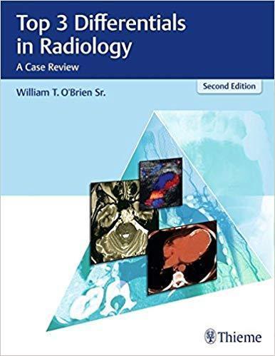
3 مورد برتر در رادیولوژی: بررسی موارد
خالی
مشخصات کتاب
ISBN (شابک): | 978-1626232785 |
قطع: | رحلی |
ناشر: | |
تعداد صفحات: | 720 |
سال و نوبت چاپ: | 2nd Edition-2018 |
نوع جلد: | هارد |
قیمت خرید کتاب : 1,299,200 تومان
جزییات بیشتر :
This fully revised second edition of Top 3 Differentials in Radiology provides a comprehensive core exam review of frequently encountered imaging gamuts in all major radiological subspecialties. Author William O'Brien utilizes the widely acclaimed format of his first edition, with 330 new and updated radiology cases organized into 12 core subspecialty sections. The last section, "Roentgen Classics," includes cases from each of the previous core sections, with imaging findings characteristic of a single diagnosis.
This book reflects content included in the current radiology board examinations with accurate and concise descriptions of key differentials, which are integral to acing the boards. Each case is formatted as a two-page unit. The left page features clinical images, succinctly captioned radiographic findings, and pertinent clinical history. The right page includes the key imaging gamut, differential diagnoses rank-ordered by the Top 3, additional diagnostic considerations, and clinical pearls.
Key Features
- More than 700 high quality images, including advanced imaging techniques
- Rank ordered differentials organized into the Top 3 and additional diagnostic considerations
- A high-yield review of important imaging and clinical manifestations for all entities on the list of differentials
- Imaging pearls at the end of each case provide a quick review of key points
This outstanding resource provides radiologists and trainees with a solid foundation of core radiology topics and a wide spectrum of key imaging findings encountered in clinical practice. It is a must-have for radiology residents preparing for board examinations. Veteran radiologists looking for a comprehensive review of critical topics in radiology will also find this book invaluable.
این کتاب مطالب مندرج در کنکور هیئت رادیولوژی فعلی را با شرح دقیق و مختصر از دیفرانسیل های کلیدی ، که برای acing تابلوها ضروری است ، منعکس می کند. هر پرونده به عنوان یک واحد دو صفحه ای قالب بندی می شود. در صفحه سمت چپ تصاویر بالینی ، یافته های رادیوگرافی به صورت موجز و تاریخچه بالینی مربوط قرار دارد. صفحه سمت راست شامل طیف تصویربرداری کلیدی ، تشخیص افتراقی مرتب شده توسط 3 صفحه ، ملاحظات تشخیصی اضافی و مرواریدهای بالینی است.
ویژگی های اصلی
بیش از 700 تصویر با کیفیت بالا ، از جمله تکنیک های تصویربرداری پیشرفته
مرتباً دستورالعمل های ترتیب یافته به 3 مورد برتر و ملاحظات تشخیصی اضافی را ترتیب دهید
مروری بر بازده تصویربرداری مهم و تظاهرات بالینی برای همه افراد موجود در لیست دیفرانسیلها
تصویربرداری از مرواریدها در پایان هر مورد ، بررسی سریع نکات کلیدی را ارائه می دهد
این منبع برجسته ، رادیولوژیست ها و کارآموزان را با پایه ای کامل از مباحث رادیولوژی هسته ای و طیف گسترده ای از یافته های تصویربرداری کلیدی که در عمل بالینی مشاهده می شود ، فراهم می کند. لازم است که ساکنین رادیولوژی برای معاینات هیئت مدیره آماده شوند. رادیولوژیست های جانباز که به دنبال بررسی جامع مباحث مهم رادیولوژی هستند نیز این کتاب را ارزشمند می دانند.
بازدید : 2167 مرتبه
محصولات مشابه
ناشرین
Elsevier (664) LWW (141) Springer (113) McGraw-Hill Education (106) تیمورزاده نوین (80) Thieme (68) Wiley-Blackwell (62) CRC Press (57) Academic Press (46) Oxford University Press (38) Cambridge University Press (32) Kaplan Publishing (24) Saunders (24) McGraw Hill / Medical (22) Elsevier (18) American Academy of Ophthalmology (17) Mosby (16) Thieme (16) Jaypee (15) Jones & Bartlett Learning (15) Pearson (12) American Academy (12)

