جزییات کتاب Imaging Anatomy: Text and Atlas Volume 3: Bones, Joints, Muscles, Vessels, and Nerves 2023

طبقه : آناتومی

آناتومی تصویربرداری: متن و اطلس جلد 3: استخوان ها، مفاصل، ماهیچه ها، عروق و اعصاب

Imaging Anatomy: Text and Atlas Volume 3: Bones, Joints, Muscles, Vessels, and Nerves 2023
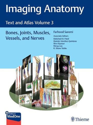
Imaging Anatomy: Text and Atlas Volume 3: Bones, Joints, Muscles, Vessels, and Nerves 2023 - آناتومی

مشخصات کتاب

ISBN (شابک):

978-1626239845

قطع:

رحلی

ناشر:

Thieme Medical Publishers Inc

تعداد صفحات:

960 pages

سال و نوبت چاپ:

1st ed. 2023 edition

نوع جلد:

هارد_سیمی

قیمت خرید کتاب : 1,999,200 تومان

قیمت خرید کتاب : 1,400,000 تومان

قیمت خرید نرم افزار : 200,000 تومان

جزییات بیشتر :

An in-depth guide to upper and lower extremity anatomy based on the latest imaging techniques

While the study of anatomy plays a fundamental role in the practice of medicine, most textbooks don’t rely on modern imaging and post-processing methods to depict and increase its understanding. Imaging Anatomy Text and Atlas Volume 3: Bones, Joints, Muscles, Vessels, and Nerves is the third in a series of four richly illustrated radiologic references edited by distinguished radiologist Farhood Saremi. The atlas is coedited by esteemed colleagues Dakshesh B. Patel, Damián Sánchez-Quintana, Hiro Kiyosue, Meng Law, and R. Shane Tubbs and features contributions from an impressive group of international experts. The succinctly written text and superb images fill a gap in the literature, with descriptions of relevant anatomical components in the context of current advances in imaging technology and science.

This exquisitely crafted atlas combines fundamental core anatomy principles with modern imaging and post-processing methods to increase understanding of intricate anatomical features. Twenty-four concise chapters cover terminology and classification of musculoskeletal structure, bones, muscles, joints, arteries, veins, nerves, and lymphatics. High-quality dissecting imaging anatomy, discussion of anatomical variants, postsurgical anatomy, and important pathology examples provide a strong foundation for differentiating normal versus pathologic anatomy.

Key Highlights

  • State-of-the-art CT, MR, angiography, and ultrasound techniques infused with 3D reformations, color coded volume rendering, and 3-7 Tesla MR views delineate anatomy in great detail
  • Cross-sectional and topographic cadaveric views and illustrations by world-renowned anatomists improve the ability to grasp difficult radiology concepts
  • Consistently formatted chapters including an introduction, embryology, review of anatomy, discussion of anatomical variants, surgical anatomy, and congenital and acquired pathologies enhance learning

This unique atlas provides a virtual, user-friendly dissection experience, making it a must-have reference for medical students, radiology residents and veteran radiologists, internists, and general surgeons, as well as vascular and transplant surgeons. 

This book includes complimentary access to a digital copy on https://medone.thieme.com.

Publisher's Note: Products purchased from Third Party sellers are not guaranteed by the publisher for quality, authenticity, or access to any online entitlements included with the product.

راهنمای عمیق آناتومی اندام فوقانی و تحتانی بر اساس آخرین تکنیک های تصویربرداری

در حالی که مطالعه آناتومی نقش اساسی در عمل پزشکی ایفا می کند، اکثر کتاب های درسی برای به تصویر کشیدن و افزایش درک آن به روش های تصویربرداری مدرن و پس پردازش تکیه نمی کنند. متن تصویربرداری آناتومی و اطلس جلد 3: استخوان‌ها، مفاصل، ماهیچه‌ها، عروق و اعصاب سومین مورد از سری چهار مرجع رادیولوژیکی با مصور غنی است که توسط رادیولوژیست برجسته فرهود صارمی ویرایش شده است. این اطلس توسط همکاران محترم داکشش بی پاتل، دامیان سانچز-کوینتانا، هیرو کیوسو، منگ لاو، و آر. شین تابز تدوین شده است و شامل مشارکت های گروهی چشمگیر از کارشناسان بین المللی است. متن مختصر نوشته شده و تصاویر عالی، شکافی را در ادبیات، با توصیف اجزای تشریحی مرتبط در زمینه پیشرفت‌های فعلی در فناوری تصویربرداری و علم، پر می‌کند.

این اطلس که به شکلی عالی ساخته شده است، اصول بنیادی آناتومی هسته ای را با روش های تصویربرداری مدرن و پس پردازش ترکیب می کند تا درک ویژگی های آناتومیک پیچیده را افزایش دهد. بیست و چهار فصل مختصر اصطلاحات و طبقه بندی ساختار اسکلتی عضلانی، استخوان ها، ماهیچه ها، مفاصل، شریان ها، وریدها، اعصاب و لنفاوی را پوشش می دهد. آناتومی تصویربرداری کالبد شکافی با کیفیت بالا، بحث در مورد انواع آناتومیک، آناتومی پس از جراحی، و نمونه‌های مهم پاتولوژی، پایه‌ای قوی برای افتراق آناتومی طبیعی از پاتولوژیک فراهم می‌کند.

نکات برجسته کلیدی

     تکنیک های پیشرفته سی تی، ام آر، آنژیوگرافی و اولتراسوند با اصلاحات سه بعدی، رندر حجمی کدگذاری شده رنگی، و نماهای MR 3 تا 7 تسلا، آناتومی را با جزئیات بسیار مشخص می کند.
     نماها و تصاویر جسد مقطعی و توپوگرافی توسط آناتومیست های مشهور جهان توانایی درک مفاهیم دشوار رادیولوژی را بهبود می بخشد.
     فصل‌هایی که به طور پیوسته فرم‌بندی شده‌اند، شامل مقدمه، جنین‌شناسی، بررسی آناتومی، بحث در مورد انواع آناتومیک، آناتومی جراحی، و آسیب‌شناسی مادرزادی و اکتسابی یادگیری را افزایش می‌دهند.

این اطلس منحصربه‌فرد یک تجربه تشریح مجازی و کاربرپسند را فراهم می‌کند و آن را به یک مرجع ضروری برای دانشجویان پزشکی، دستیاران رادیولوژی و رادیولوژیست‌های کهنه کار، متخصصین داخلی و جراحان عمومی و همچنین جراحان عروق و پیوند تبدیل می‌کند.

این کتاب شامل دسترسی رایگان به یک نسخه دیجیتال در https://medone.thieme.com است.

توجه ناشر: محصولات خریداری شده از فروشندگان شخص ثالث توسط ناشر برای کیفیت، اصالت یا دسترسی به حقوق آنلاین موجود با محصول تضمین نمی شود.


خريد کتاب پزشکي با تخفيف, usmle step 1 books, usmle step 2 books, usmle step 3 books, usmle, usmleiran, usmle ایران, مرکز تخصصی usmle در ایران, 2021, 2022, امتحانات کانادا و استراليا و امريکا , آزمون هاي پزشکي خارج از کشور, مديکال بوک, کتاب رفرنس پزشکي, دانلود کتاب پزشکي, انتشارات کتب پزشکي, انتشارات پزشکي

بازدید : 231 مرتبه

محصولات مشابه